Medical imaging
My contribution
I implemented a 3D U-Net training and inference workflow and a separate pipeline for leakage-controlled full-volume evaluation.
Research implementation · validation limitations documented
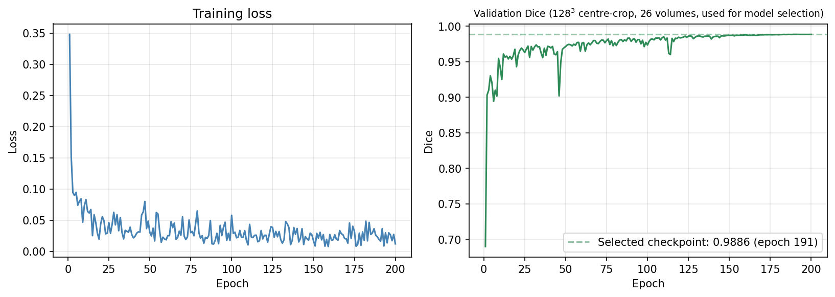
Dice 0.9886 on centre-cropped model-selection volumes; independent full-volume evaluation is pending.
Problem
Automatic segmentation of liver structures from abdominal CT volumes for downstream clinical and computational analysis.
Approach
A 3D U-Net uses the MSD Task03 liver dataset (131 labelled CT cases, including the 26-volume model-selection split), with foreground-biased patch sampling, a combined soft-Dice + BCE loss, cosine learning-rate annealing, and Gaussian sliding-window inference.
Result
Historical result: 0.9886 Dice on the 26-volume 128³ centre-cropped validation split used for model selection, after 200 epochs on a single Kaggle T4 GPU. The trained checkpoint has since been lost, so this value cannot be re-evaluated.
Limitations
This is not an independent or full-volume test result, and it cannot be reproduced because the checkpoint was lost. No boundary-distance metric is reported for this model: the historical HD95 implementation was invalid and cannot be recomputed. A leakage-controlled full-volume evaluation pipeline with physical-unit HD95 has been implemented; independent evaluation is pending retraining on the original NIfTI data.